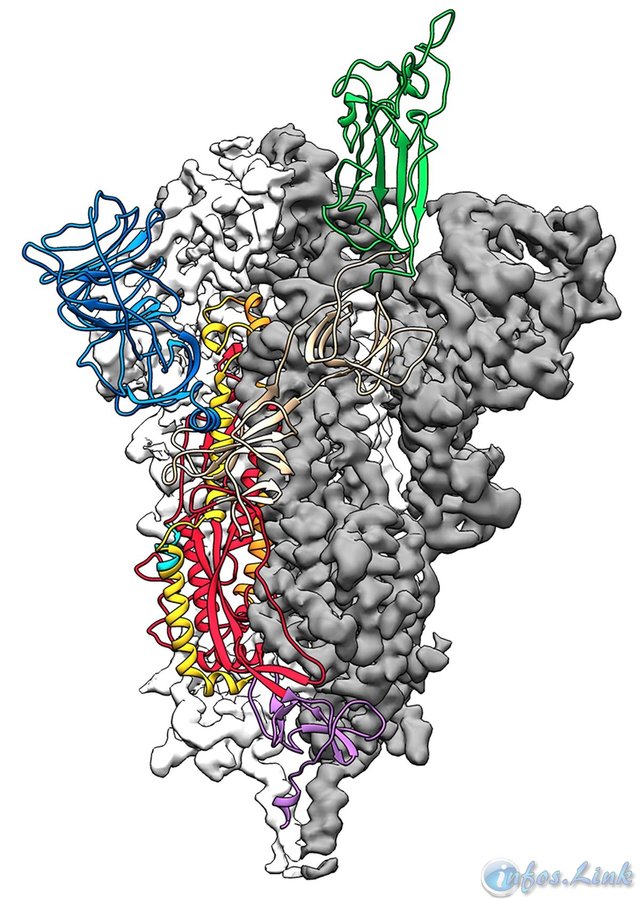
Scientists publish molecular map of new coronavirus

American scientists published Wednesday in the journal Science the first three-dimensional atomic map of the part of the new coronavirus that infects human cells, an important step in the development of treatments and vaccines.
A team of researchers from the University of Texas at Austin and the National Institutes of Health (NIH) used a technology called cryo-electron microscopy (distinguished by the Nobel Prize in chemistry in 2017) in order to model the part of the virus that attaches to cells, spikes called spicule proteins.
"The tip is the antigen that we would like to introduce into humans to preemptively trigger the production of antibodies by the immune system, so that it is ready to respond to an attack the day the real virus arrives," explains AFP Jason McLellan, the scientist who conducted the study.
He and his team have been studying other viruses in the same family for years, including SARS and Mers.
Drawing on this experience, and based on the genome published by the Chinese at the start of the epidemic, they made a stable version of the spikes of the virus in the laboratory.
Their molecular structure is now available to researchers around the world.
"It's a beautiful, clean structure of one of the most important proteins in the coronavirus, a real breakthrough in understanding how this coronavirus finds and penetrates cells," comments virologist Benjamin Neuman, at Texas A&M University-Texarkana, who n did not participate in this work.
The card should notably help researchers understand how the virus hides and how to neutralize it, giving possible recipes for antiviral drugs, for people already infected, and a vaccine.