Stuff under a microscope (and my way of introducing myself)
Hello Steemit Community!
My name is Andrea and I live in Venezuela, a medium-sized country in South America. Here, I work as a Bioanalyst. You might wonder what a bioanalyst does. Well, the answer is not simple at all, because we analyze everything from blood, tissues, urine, feces and body fluids.
So in my first post I bring you a selection of things that I have seen through the microscope while I work.
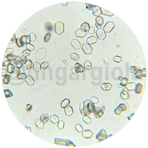
- Image: Uric acid crystals (400x).
- Sample: urine.
Your body produces uric acid when it breaks down purines —substances that are found naturally in your body—. Yeah, they can be beautiful (but painful sometimes).
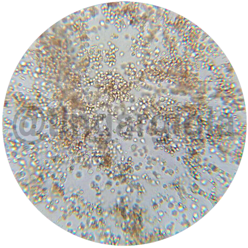
- Image: Hematuria and pyuria (400x).
- Sample: urine.
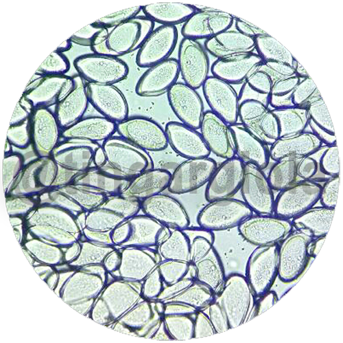
- Image: Enterobius vermicularis eggs (400x)
- Method: Graham.
Also known commonly as pinworm is the most common intestinal parasite. It is a nematode that inhabits the human terminal ileum, colon and appendix.
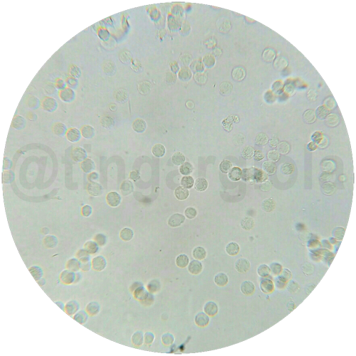
- Image: leukocytes 400x.
- Sample: urine.
The presence of leukocytes in the urine could be a sign that you have an infection or an obstruction in the urinary tract or bladder (ouch!).
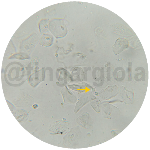
- Image: Squamous epithelial cells (400x).
- Yellow arrow: leukocyte.
- Sample: urine.
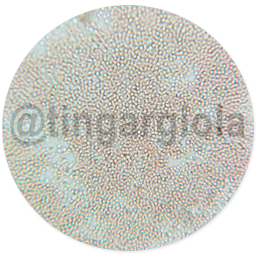
- Image: Hematuria (400x).
- Sample: urine.
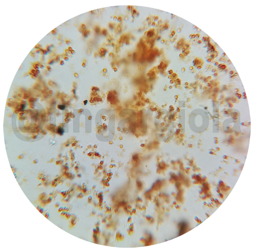
- Image: Leukocytes and erythrocytes result of infection by Entamoeba histolytica (400x).
- Sample: feces.
Entamoeba histolytica is well recognized as a pathogenic ameba, associated with intestinal and extraintestinal infections.
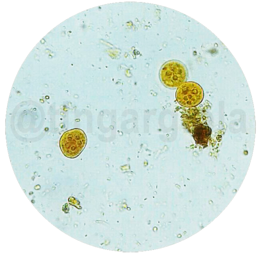
- Image: Entamoeba coli cysts (400x).
- Sample: feces.
Entamoeba coli is generally considered nonpathogenic and reside in the large intestine of the human host. Tip: they can have between 1-8 nuclei.
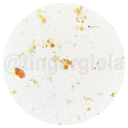
- Image: Entamoeba histolytica/dispar cyst (400x).
- Sample: feces.
Infections caused by the Entamoeba histolytica/Entamoeba dispar complex are of cosmopolitan distribution. E. histolytica is known as the pathogenic specie of the complex, while E. dispar is considered commensal (non-pathogenic). They can be differentiated through Polymerase Chain Reaction (PCR)
Extra
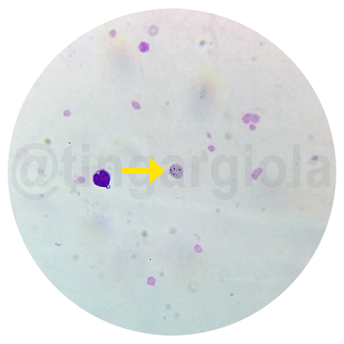
Image: (yellow arrow) Babesia spp. merozoite found in the peripheral blood of a puppy (1000x).
Method: Buffy coat smear.
Sample: peripheral blood.
Babesia is a genus of protist parasites that cause babesiosis in animals and humans. Babesia has the ability to infect red blood cells, where it carries out its life cycle, resulting in lysis of red blood cells and therefore, severe anemia. Tip: They spread by certain ticks.
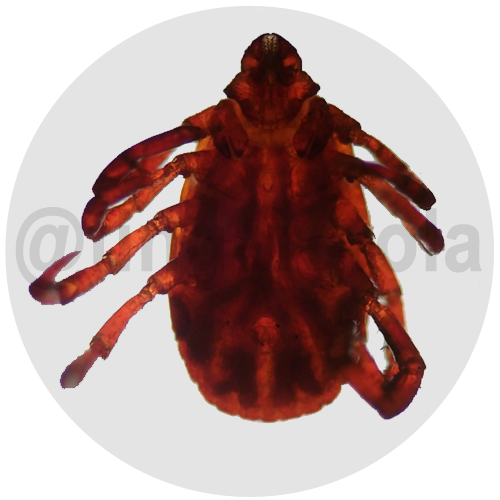
Image: Tick (40x).
welcome here and enjoy Steemit! :)
Welcome to steemit @tingargiola. Join #minnowsupportproject for more help. @OriginalWorks.
Thank you so much! It's wonderful to be in Steemit!
Congratulations @tingargiola! You have completed some achievement on Steemit and have been rewarded with new badge(s) :
Click on any badge to view your own Board of Honor on SteemitBoard.
For more information about SteemitBoard, click here
If you no longer want to receive notifications, reply to this comment with the word
STOPWelcome to Steemit, Andrea. Tough job! Thank you for sharing these samples with us. They look so different than what they are under the microscope.
Amazing how colourful and artistic those samples are!! Nice to see you in here.. and good luck with future postings.
Thanks @gwelton! Yeah, they are very artistic at their own way :D. Greetings!
Good posting, welcome to steemit @tingargiola
Look who's here ^^ @Tingargiola, In case this has been your first Introdusemyself Post i'm here to welcome you to Steemit. I hope you have a lot of fun here and you may follow me. Have a great time @rightuppercorner
Thanks @rightuppercorner!
We want more pics like this :D
Welcome to Steemit @tingergiola. Wow, this is a very unique introduction post. Thanks for sharing.
That's so sweet, @spectrumecons! Thanks!
Hi Andrea... Nice to meet you... Welcome to Steemit..🌹.. This is a nice and very different type of intro post... I hope you will do great over here as a steemian..😊.. Follow Me @onority