SENSE ORGAN - THE GIFT OF SIGHT
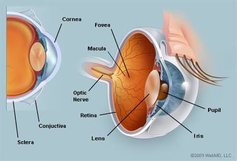
The eyes are located in a bony socket in the skull called the eye socket. The eyeballs are connected to the eye socket by the occular muscles. The eyes are made up of three layers: choroid, retina and sclerotic. The eyes are one of the sense organ in the body that is most highly developed. The eye uses the pupil to regulate the amount of light entering the eye. The eyeballs are globular in shape with a diameter of 24mm. The eye contains a fluid that helps to bathe the tissues of the pupil, cornea( contains a fluid called aqueous humour), and lens.
.jpg) source
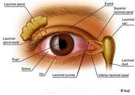
sourceThere are photoreceptors present in the eye called rods and cones that help to detect the presence of light. The cones are sensitive to bright light and detect colours. This is due to a chemical substance called iodospin. The rods are sensitive to light of low intensity. They contain a sensitive pigment called rhodospin. The rhodospin is made up of protein and vitamin A. When the rhodospin receives light rays chemical reaction takes place which releases a yellow pigment called retinene and energy that helps to stimulate the optic nerve to carry the light rays to the brain. The brain interprets it as light. Vitamin A also help to regenerate rhodospin when the eyes are closed. That's why deficiency of vitamin A causes night blindness. The eyes also contains a fluid called the vitreous humour. Both the vitreous humour and the aqueous humour are nutritive fluids and are the only source of nutrients to the cornea, iris and lens. This fluids help to refract the light and produce an image on the retina. There is a small depression in the retina that helps to give an accurate interpretation of an image. This small depression is called the fovea. Iris are pigmented blood vessels that determine what is usually called the "colour of the eye". The tears gland secretes a solution of sodium hydrogencarbonate and sodium chloride to moisten the surface of the cornea and conjunctiva. The tears gland are called the larcrymal gland. They produces tears that contains lysozyme which destroy bacteria's. The choriod are network of blood vessels that supply food and oxygen to the eye. There is a region where there are no light-sensitive cells. This is where the nerve fibres leave the eye to enter the optic nerve. If any part of an image falls on this region, nothing will be recorded in the brain. This is called the blind spot.
Eye movement :- Both eye's can move on their sockets by the help of extrinsic voluntary eye muscles. Both eyes are not independent of eachother. They must move together.
Colour vision :- We have three types of cones that are sensitive to red, blue, and green light respectively. These cones are stimulated by their own particular wavelength of light so that, according to the kind of cell and the numbers stimulated, the brain receives an impression of colour. Only primates among mammals appreciate colour. The others See's only black, white or shades of grey.
PROTECTION OF THE EYE'S
The eyelid moisten the conjunctiva and also prevent bright light from damaging the eye. The eye lash helps to prevent dust from entering the eyes. The Bony socket protects the eye's from damage. The tears gland destroy bacteria or germs.
Accommodation :- This is the ability of the eye to alter its focal length is called accommodation. Images made on the retina are upside down and laterally inverted. For this image to be seen properly the brain has to correct this image so that the object can be seen properly. To get a clear on the retina, focusing is needed. It is brought about by the contraction and relaxation of the cilliary muscles. The cilliary muscles contains circular and radial muscles.
TYPES OF EYE MOVEMENT
1.) Conjugate movement :- This is a movement of both eyeballs in the same direction. It is due to contraction of medial rectus of one eye and lateral rectus of the other eye.
2.) Disjugate movement :- both eyeball's move in opposite direction. Disjugate movement is one of two types. The convergence (both eyeballs move towards the nose) and divergence (both eyeball's move towards temporal side.
3.) Saccadic movement :- This is the quick movement of both eyeballs when its shifted from one object to another.
4.) Pursuit movement :- This is when the eyeballs follow a moving object.
.jpg) source
sourceEYE DEFECTS
1.)long sightedness :- This is a kind of defects whereby someone cannot see near object clearly but it can see distant object clearly. It is occurs when the eyeball is too short and when parrallel rays of light from near objects focus behind the retina. Long sightedness is corrected using aconvex lens.
2.) Short sightedness :- This is a kind of eye defects where the person cannot see far objects focused in front of the retina. It is caused when the eyeball is too long and when parallel rays light from far object focus at the front of the retina. It can be corrected using a concave lens.
3.) Presbyopia :- its a kind of eye defects at old age caused as a result of loss of elasticity of the lens and cilliary muscle. It can be corrected using a bifocal lens.
4.) Astigmatism :- This is an eye defects causes by unevenly bending of light ray at the cornea. Someone suffering from this cannot focus objects horizontally and vertically. It is corrected by wearing glasses with cylindrical lens.
.jpg) source
sourceCARE OF THE EYE
1.) Always use clean handkerchief or clean water to clean the eyes.
2.) Eat food that contains vitamin A.
3.) Do not read with dim light or direct sunlight or sitting very close to a TV.
4.) Do not use glasses that are not recommended.
5.) Do not rub the eyes with dirty fingers when removing foreign bodies.